What Is a Laparoscopic Ileocolectomy and Right Colectomy?
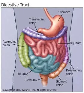
A laparoscopic ileocolectomy is an operation that removes a diseased section of the ileum (last segment of the small bowel) and ascending colon.
In a right colectomy, the surgeon removes the ascending colon, but leaves the ileum. Both surgeries are used to treat the following:
- Cancer
- Noncancerous growths
- Areas of swelling (inflammation) caused by Crohn's disease
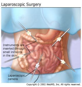
The term "laparoscopic" refers to a type of surgery called laparoscopy. Laparoscopy enables the surgeon to operate through very small "keyhole" incisions in the abdomen. A laparoscope, a small, lighted tube with an attached camera, is placed through a small incision near the bellybutton. (In some cases, these incisions may not be near the bellybutton.)
What Happens During an Ileocolectomy and Colectomy?
There are four main steps to these surgeries.
Step 1: Positioning the Laparoscope
Once you are under anesthesia, the surgeon will make a small cut (about 1/2 inch) often near the bellybutton. A laparoscope will be inserted into the abdomen through this incision. Images taken by the laparoscope will be projected onto video monitors placed near the operating table.
Once the laparoscope is in place, the surgeon will make five or six more small incisions in the abdomen. Surgical instruments will be placed through these incisions to complete the surgery.
Step 2: Freeing the Bowel
In the ileocolectomy, the diseased section of the ileum and ascending colon must be cut away from the healthy bowel. Before this section can be removed, it must be freed from its supporting structures. In the right colectomy, the ascending colon must be freed from its supporting structure.
The bowel is attached to the abdominal wall by a layer of tissue called the "mesentery." The mesentery also contains the main blood vessels (arteries) that supply blood to the ileum and ascending colon. These arteries will be carefully cut and closed. In an ileocolectomy, the surgeon will then free the ileum and ascending colon from the mesentery. In a right colectomy, only the ascending colon will be freed from the mesentery. After the bowel is free from the mesentery, the surgeon will cut away the diseased section of bowel.
Step 3: Removing the Diseased Bowel
Because the incisions used in laparoscopy are very small, the diseased section of bowel must be removed in a special way. Your surgeon will enlarge one of the incisions and place a bag into the abdominal cavity. The diseased bowel is placed into this bag. The bag is then pulled out of the enlarged incision.
Step 4: Rejoining the Ends of the Colon
After the bag has been removed, the ends of the colon will be pulled through the enlarged incision. Your surgeon will then use a stapling device or sutures (stitches) to rejoin the bowel. This rejoining is called an "anastomosis."
Before the operation is completed, the surgeon will rinse out the abdominal cavity and check the anastomosis for leaks. Lastly, all of the incisions in the abdomen will be stitched or taped closed.
Recovering at Home
You will be encouraged to increase your activity level steadily once you are home. Walking is great exercise! Walking will help your general recovery by strengthening your muscles, keeping your blood circulating to prevent blood clots, and helping your lungs remain clear. If you are fit and did regular exercise before surgery, you may resume exercising when you feel comfortable and your doctor gives the approval. However, strenuous exercise, heavy lifting, and abdominal exercises such as sit-ups should be avoided for six weeks after the surgery.
You will be sent home on a soft diet, which means you can eat most everything except raw fruits and vegetables. You should continue this diet until your post-surgical check-up. If the diet is making you constipated, please call your doctor for advice.