1 of 4 / Overview
View All
What You Need to Know About Infertility
Infertility doesn’t mean you can never have a child.
Effects of Exercise, Age, and Weight on Fertility
All three can affect fertility. Find out how.
Your Guide to Infertility in Women
Learn the causes of female infertility.
Luteal Phase Defect
A luteal phase defect is a disruption in a woman's monthly menstrual cycle.
2 of 4 / Symptoms
View All
3 of 4 / Tests & Diagnosis
View All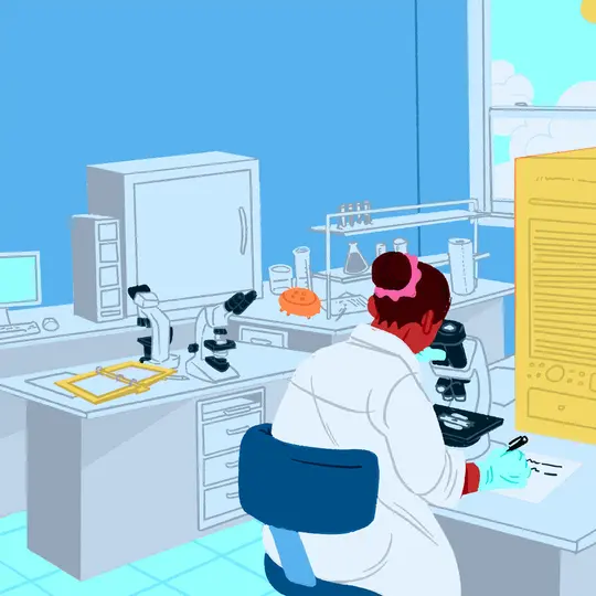
4 of 4 / Treatment
View All
In Vitro Fertilization
How much does it cost? How often does it succeed? Find these and other details.
Artificial Insemination
This simple procedure can be a good initial treatment for infertility.
Using an Egg Donor
This increasingly common procedure has a high success rate.
Surrogate Mothers
Surrogacy is still somewhat controversial. But it is one more option people have for having a baby through new technologies.
Suggested Reads about Infertility and Reproduction
Sperm Donors May Not Be as Anonymous as They Think
Sperm donors aren’t always anonymous anymore. Read about how DNA testing and tech are upending sperm bank donation anonymity.
COVID Could Impair Men’s Sperm for Months: Study
A COVID infection can reduce sperm count and hinder the ability of sperm to swim for at least 3 months, according to European researchers.
AI Meets Embryos: The Future of IVF
Conceiving a child in the next five to 10 years might look like something out of a science fiction movie.
Scientists Create 'Vagina on a Chip': What to Know
A microfluidic chip designed to mimic the vagina's microbiome is likely to drive vital new treatments in the long-neglected field of women's reproductive health.
Top Search Terms for Infertility & Reproduction
8 million+ Physician Ratings & Reviews
Find Doctors and Dentists Near You
You can also search by physician, practice, or hospital name