Mammography uses special X-ray images to spot abnormal growths or changes in breast tissue.
Using a digital X-ray machine made especially for breast tissue, a technician takes pictures from at least two angles to make a set of images for each of your breasts. This set of images is called a mammogram. Breast tissue looks white and opaque, and fatty tissue appears darker and translucent.
Why Do I Need a Mammogram?
Mammograms are done as part of a regular physical exam to provide a baseline reference or to check any unusual changes in the breast. It’s the best screening test for lowering the risk of dying from breast cancer.
A mammogram can help your health care provider decide if a lump, growth, or change in your breast needs more testing. The mammogram also looks for lumps that are too small to feel during a physical exam.
Why Should I Get a Mammogram?
Mammography can be your best defense against breast cancer because it can often spot the disease in its early stages, before it can be felt during a breast exam. Research shows that mammography can increase breast cancer survival.
How Should I Prepare for a Mammogram?
You can eat, drink, and take medications as usual.
Tell your doctor or the technician if you are pregnant or think you may be pregnant.
Don’t wear body powder, cream, deodorant, or lotion on your chest the day of the test. They may interfere with the X-rays.
You’ll be asked to remove all jewelry and clothing above the waist and put on a hospital gown. You may want to wear a two-piece outfit the day of the test.
What Happens During a Mammogram?
Registered mammography technologists perform the test. Most of them are women. A doctor specialized in interpreting imaging studies (radiologist) will interpret the X-rays.
You’ll stand in front of an X-ray machine. The technologist will put your breast between two radiographic breast supports. The supports will be pressed together, gently flattening the breast. This is necessary to get the clearest possible picture with the least amount of radiation. You may feel some discomfort or slight pain, but it will last for only a few seconds while the X-ray is taken. If you feel that there’s too much pressure on your breast, tell the technologist.
You may want to schedule your appointment 7 to 10 days after the start of your period, when your breasts are least likely to be tender.
The breast will be imaged in several positions so the radiologist can see all the tissue. For a routine breast screening, two pictures are taken of each breast. This exam takes about 20 minutes. Many centers also do 3D mammography. This involves many more pictures of the breast taken at various angles to make a 3D picture.
After looking at the digital images, the radiologist may ask for more images or a breast ultrasound for a more accurate diagnosis.
What Happens After a Mammogram?
You may have temporary changes in skin color or mild aching because of the compression. You can take aspirin or ibuprofen to ease the discomfort. Generally, you can go back to your regular activities right away.
Your doctor will get the results of your mammogram. They’ll speak with you about what the test results could mean and what other tests you might need.
All mammography facilities are required to send your results to you by mail within 30 days. You’ll be contacted within 5 business days if there is a problem with your mammogram. If you don’t hear about your test findings within 10 working days, call your doctor.
According to the American Cancer Society, about one or two mammograms out of every 1,000 lead to a diagnosis of cancer. About 10% of women will need more mammography. Don't be alarmed if this happens to you. Only 8% to 10% of those women will need a biopsy, and 80% of those biopsies won't be cancer. Those odds may improve with more widespread use of 3D mammography.
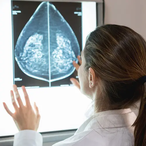
What Does a Suspicious Area Look Like on a Mammogram?
- A lump or mass with a smooth, well-defined border usually isn’t cancerous. An ultrasound can look inside the lump. If it’s filled with fluid, it’s called a cyst, and it’s usually not cancer, but your doctor might recommend a biopsy.
- A lump that has an irregular border or a starburst appearance raises more concern. A biopsy is usually recommended.
- Deposits of calcium (calcifications) can be large or small, and they might or might not be cancerous. If the deposits are very small, you may need more tests and a biopsy.
What Is a Diagnostic Mammogram?
In a screening mammogram, the breast is X-rayed from top to bottom and from side to side. A diagnostic mammogram focuses on a particular lump or area of abnormal tissue.
You might have a diagnostic mammogram after a screening mammogram spotted something unusual. Or your doctor might recommend one first, without a screening mammogram, if you have symptoms they'd like to check further.
How Well Do Mammograms Work?
These imaging tests help doctors diagnose about 75% to 85% of breast cancers. Detection rates get better as a woman ages, because breasts become less dense with age. This makes tissue easier to see through on mammograms.
Advancing technology raises detection rates. One study showed that using 3D mammography along with digital mammograms improved detection rates and lowered the number of women who had to return for more tests because of a suspicious finding.
How Often Should I Have a Mammogram?
Your risk of breast cancer goes up as you age. But experts disagree about when you should have your first mammogram.
The American Cancer Society recommends that women ages 40 to 44 should have a choice to start yearly screening mammograms. Women 45 to 54 should have a mammogram each year, and those 55 years and over should get mammograms every 1 to 2 years. But the U.S. Preventive Services Task Force recommends screening every 2 years from ages 50 through 74 and says the decision to start yearly screening mammograms before age 50 should be an individual one. Talk with your doctor about when you should start getting them.
If your doctor tells you you're at high risk for breast cancer, or if you have close family members who got the disease at an early age, you might want to consider getting screened earlier.
Most experts recommend that you continue to have these screenings as long as you are in good health and are expected to live at least another 10 years.
Mammograms are an important part of your health history. If you go to another health care provider or move, take the film (mammogram) with you.
Should I still have clinical breast exams done?
Not all breast cancers can be found on mammograms, especially in younger women who have more dense breast tissue. You may also have breast exams done by your health care provider (physician or nurse) every 3 years starting at age 20 and every year starting at age 40.