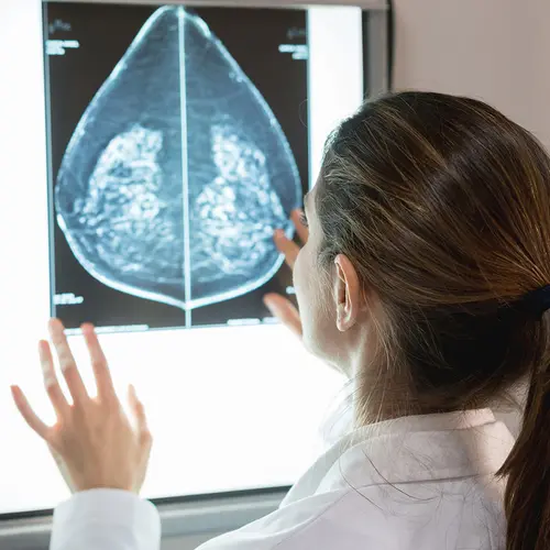
What Is a Breast Biopsy?
A breast biopsy is a procedure in which your doctor takes out cells or a small piece of tissue from part of your breast. They look at it under a microscope for signs of cancer. It’s the only way to know for sure if a possible trouble spot is cancer.
Why Is a Breast Biopsy Done?
If your doctor finds something suspicious during a routine breast exam, mammogram, or ultrasound, they may recommend this test. Possible signs of trouble include:
- A lump or mass that you can feel in your breast
- Masses filled with fluid (cysts) or small calcium deposits (microcalcifications)
- Nipple problems like bloody discharge
Types of Breast Biopsy Procedures
Your doctor will recommend a breast biopsy procedure based on things like:
- The size of the lump or suspicious area
- Where it is
- Whether there’s more than one unusual area
- If you have other medical problems
- What you prefer
Your doctor may take a sample of tissue through surgery or through a procedure called a minimally invasive biopsy. Minimally invasive procedures offer:
- Less scarring
- Less pain
- Lower risk of infection
- Possibly lower hospital costs
- Shorter recovery time
- Return to typical daily activities right away
Common minimally invasive biopsy procedures include:
- Fine-needle aspiration. Your doctor uses a small needle to take a sample of cells from the area in question. If the lump is a cyst (a fluid-filled sac), the procedure may cause it to collapse. This fluid will be looked at under a microscope for any signs of cancer. If the lump is solid, cells can be smeared onto slides for examination.
- Ultrasound-guided core biopsy. Your doctor puts a needle into the breast tissue. Ultrasound helps confirm the exact location of the potential trouble spot so the needle goes to the right place. Tissue samples are then taken through the needle. Ultrasound can see the difference between cysts and solid lesions.
- Vacuum-assisted breast biopsy. A suction device gets more fluid and cells through the needle. It can cut down on the number of times the needle needs to be inserted to get samples.
- Stereotactic biopsy. The medical team centers the area to be tested in the window of a specially designed instrument. Mammogram films called SCOUT films are taken so a specialist called a radiologist can examine the area to be biopsied. After using medicine to numb the area, the radiologist makes a small opening in your skin. They put a needle into the breast tissue, and computerized pictures help confirm the exact placement. Tissue samples are taken through the needle. It's common for medical professionals to take multiple tissue samples (about three to five).
Surgical procedures include:
- Open excisional biopsy. This is surgery to remove an entire lump. The tissue is then studied under a microscope. If your doctor takes a section of normal breast tissue all the way around a lump (called a lumpectomy), the biopsy is also considered a breast cancer treatment. In this technique, they may put a wire through a needle into the area to be biopsied. An X-ray helps make sure it’s in the right place, and a small hook at the end of the wire keeps it in position. The surgeon uses this wire as a guide to find the suspicious tissue.
- Sentinel node biopsy. This method helps ensure that only the lymph nodes most likely to have cancer are removed. It pinpoints the first lymph node a tumor drains into (called the sentinel node). To spot it, your doctor puts a radioactive tracer, a blue dye, or both into the area around the tumor. That helps them figure out which lymph nodes are the first to receive drainage from the breast. These nodes would possibly be the first to be invaded by cancer cells. One to three sentinel nodes are usually removed and tested for cancer. If the sentinel node is positive, there may be other positive lymph nodes upstream. If it is negative, it is highly likely that all of the upstream nodes are negative.
You may also have an axillary node dissection. Your doctor takes out at least six of the lymph nodes under your arm and sends them to a lab to be checked for cancer. This is a very reliable way to check the extent of your cancer. But it can take longer to recover, and it can have complications like arm swelling (lymphedema) or nerve damage. It is rarely done anymore if at all avoidable.
After surgery, watch for warning signs of an infection or swelling in your arm or hand. Call your doctor right away if you notice a buildup of fluid, redness, or other symptoms of infection.
Cells or tissues that are removed are given to a pathologist, a doctor who specializes in diagnosing suspicious tissue changes.
Risks of Breast Biopsies
Breast biopsies are relatively safe. Risks include:
- Bruising
- Swelling
- Mild pain
- Bleeding
- Infection
- A change in how your breast looks, depending on how much tissue the doctor removes and how it heals
Breast Biopsy Recovery
You may need to wear a special bra and dressings over the breast biopsy site for a few days after the procedure. You’ll have small strips of tape or stitches over the place your skin was cut. Don’t try to remove these yourself. Your medical team will tell you whether someone will take them out at a later appointment or they’ll fall off by themselves.
Your team may tell you to put medicine on the biopsy area or change the bandages at home. Your doctor will give you advice on showering, bathing, and wound care.
You’ll get a prescription for pain relief if you need it, but an over-the-counter pain reliever might be enough. To lower the risk of bleeding, don’t take aspirin or products containing aspirin for the first 3 days after the procedure unless a doctor tells you to.
The area of the biopsy might be black and blue for a few days afterward, too.
Call your doctor if you notice problems like:
Breast Biopsy Results
Core needle biopsies are done with imaging so you usually confirm during the biopsy itself that the correct area was sampled. It is then marked with a clip that shows up on future mammograms or if an additional surgical biopsy is needed.
It may take several days for the pathologist to look at the sample from your biopsy and prepare a report on it. Your doctor will discuss the findings with you.
If the report says you have normal or benign (noncancerous) tissue, and your doctor still thinks the area is suspicious, you may need to have another procedure.
If the biopsy shows that you have breast cancer, the pathologist’s report will include details about the tumor. This will help your doctor recommend a treatment plan.