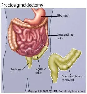
This operation removes a diseased section of the rectum and sigmoid colon. Doctors use it to treat the following conditions:
- Cancers of the colon and rectum
- Some types of noncancerous growths in the colon and rectum
- Complicated diverticulitis
The term "laparoscopic" refers to a type of surgery called laparoscopy, in which the surgeon works through very small (5-millimeter to 10-millimeter) "keyhole" cuts in the abdomen.
A laparoscope is a small telescope-like instrument. Your surgeon will use it in order to see inside you during the operation.

There are five main steps to this surgery.
1. Positioning the Laparoscope
First, you’ll get general anesthesia so that you’re “asleep.” Then your surgeon will make a small cut (about half an inch) near your belly button and place the laparoscope through it so they can see images from inside you.
Once the laparoscope is in place, the surgeon will make five or six more small (5-10 millimeter) cuts to make room for the surgical tools.
2. Dividing the Sigmoid Colon
Your surgeon will need to cut away the diseased section of your sigmoid colon and rectum. But first, they must free this section from what supports it.
The bowel is attached to the abdominal wall by a layer of tissue called the mesentery, which also contains the main blood vessels (arteries) that take blood to the left side of the colon and rectum. Your surgeon will cut and close them. Then they’ll free the sigmoid colon and part of the rectum from the mesentery, and cut away the diseased tissue. Later, they’ll remove this part of the mesentery with the diseased bowel.
In a total proctosigmoidectomy, the rectum is removed.
3. Preparing to Rejoin the Colon
The surgeon must rejoin the remaining end of the descending colon with the remaining end of the rectum.
First, they will detach a part of the healthy descending colon from the mesentery so that they can stretch it toward the rectum. They’ll also free the rectum from its mesentery so it can meet the end of the colon.
To reduce the risk of cancer cells spreading, the surgeon will wash the rectum with a special solution.
4. Removing the Diseased Bowel
The cuts used in laparoscopy are very small, so the surgeon must remove the diseased section of bowel in a special way. They will enlarge one of the cuts and place a bag into your abdominal cavity, put the diseased bowel into the bag, and then pull the bag out of the enlarged cut.
5. Rejoining the Ends of the Colon
To do this, your surgeon will use a special stapling device they place into the rectum. Doctors call this rejoining of the colon and rectum an anastomosis.
The stapling device "fires" a ring of staples to connect the two ends. The surgeon will check the anastomosis for leaks and rinse out your pelvis.
Your surgeon may place a drain into your abdomen for a few days to help you recover after surgery. And they will stitch or tape close all the surgical cuts.
Recovery
You should avoid heavy lifting and abdominal exercises such as sit-ups for 6 weeks after the surgery.
Apart from that, you should steadily build up your activity level when you get home. Walking is a great exercise choice. It will help your recovery by making you stronger, keeping your blood circulating to prevent blood clots, and helping your lungs remain clear.
Did you work out before the surgery? You can get back to exercising when you feel comfortable and your doctor says it’s OK.
When you go home, you’ll be able to eat almost everything except raw fruits and vegetables. You should continue this “soft” diet until your post-surgical check-up. If the diet makes you constipated, call your doctor's office for advice.

