What Is Retinal Vein Occlusion?

Hide Video Transcript
Video Transcript
[MUSIC PLAYING]
And in order to keep that retina healthy and perfused, there's blood vessels. There's arteries, and there's veins. And so your body can generate a thrombosis or an occlusion-- so that's a little plaque or some other reason why the blood flow through those retinal veins gets stopped. When that happens, you have a lack of perfusion and a retinal vein occlusion. So there are two ways of thinking about retinal vein occlusion, and they fall into two separate buckets.
So the first type is to base a retinal vein occlusion based on the parts of the retina that it's involving. So you can have something called a central retinal vein occlusion, and that's where you get that occlusion or thrombosis at the level of the optic nerve. And that prevents blood flow through the retinal veins all throughout the retina. So we call it a central vein occlusion because it impacts the entire retina. Or you can have something called a branch retinal vein occlusion.
And what that means is that you've only impacted a certain segment of those retinal veins. So it can either be half of the retina or a small section thereof. So the symptoms of retinal vein occlusion can vary. So the first thing is, the patient might tell me that they have a certain hemisphere of their vision that's involved or a small little scotoma, or a blank spot or a black spot. That points more to a branch retinal vein occlusion, meaning not all of their veins are involved.
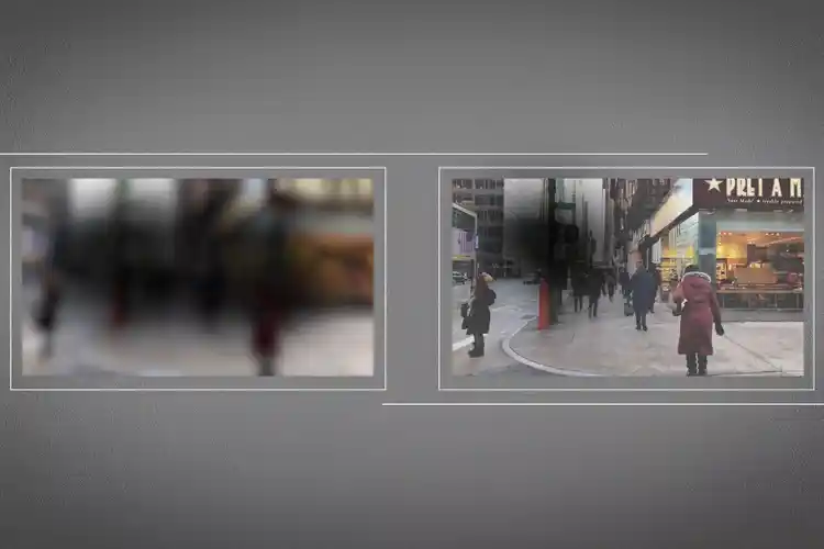
On the other hand, if a person has a central retinal vein occlusion, they're generally going to come in saying that their entire visual field is disturbed, so things look blurry or black or gray, and they're not able to see as well as they would, usually, compared to a person with a branch retinal vein occlusion. So there's a lot of things that can cause a retinal vein occlusion. And so when you have a patient who comes in with what looks like a vein occlusion, we start to go through a checklist of questions for them.
For common things, you ask them about diabetes, high blood pressure, high cholesterol. You ask them about things like a history of glaucoma, which can predispose you to developing a vein occlusion. And you also ask them about things like, "Does anyone ever tell you that you snore at night or that you have sleep apnea?" All of those things have been linked to developing a vein occlusion.
SRUTHI AREPALLI
So a retinal vein occlusion is when there is an occlusion in the blood vessels of the retina that are particularly the veins, and it causes a lack of blood flow, or a lack of perfusion, through those vessels. The way that I explain it to my patients is that if we were to look in the back of the eye, there is a structure called the retina. That's sort of like the wallpaper of the eye, and it helps us transmit a signal from the front of the eye to the optic nerve and to the brain. And in order to keep that retina healthy and perfused, there's blood vessels. There's arteries, and there's veins. And so your body can generate a thrombosis or an occlusion-- so that's a little plaque or some other reason why the blood flow through those retinal veins gets stopped. When that happens, you have a lack of perfusion and a retinal vein occlusion. So there are two ways of thinking about retinal vein occlusion, and they fall into two separate buckets.
So the first type is to base a retinal vein occlusion based on the parts of the retina that it's involving. So you can have something called a central retinal vein occlusion, and that's where you get that occlusion or thrombosis at the level of the optic nerve. And that prevents blood flow through the retinal veins all throughout the retina. So we call it a central vein occlusion because it impacts the entire retina. Or you can have something called a branch retinal vein occlusion.
And what that means is that you've only impacted a certain segment of those retinal veins. So it can either be half of the retina or a small section thereof. So the symptoms of retinal vein occlusion can vary. So the first thing is, the patient might tell me that they have a certain hemisphere of their vision that's involved or a small little scotoma, or a blank spot or a black spot. That points more to a branch retinal vein occlusion, meaning not all of their veins are involved.
On the other hand, if a person has a central retinal vein occlusion, they're generally going to come in saying that their entire visual field is disturbed, so things look blurry or black or gray, and they're not able to see as well as they would, usually, compared to a person with a branch retinal vein occlusion. So there's a lot of things that can cause a retinal vein occlusion. And so when you have a patient who comes in with what looks like a vein occlusion, we start to go through a checklist of questions for them.
For common things, you ask them about diabetes, high blood pressure, high cholesterol. You ask them about things like a history of glaucoma, which can predispose you to developing a vein occlusion. And you also ask them about things like, "Does anyone ever tell you that you snore at night or that you have sleep apnea?" All of those things have been linked to developing a vein occlusion.
