A biopsy is used to detect the presence of cancer cells in the prostate and to evaluate how aggressive cancer is likely to be. Thanks to an array of biopsy techniques and new tools to interpret the results, doctors are better able to predict when cancers are slow-growing and when they’re likely to be aggressive. That information, in turn, can help you and your doctor choose the best course of treatment.
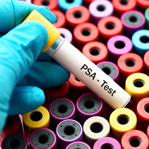
Before having a prostate biopsy performed, most men have undergone other tests for prostate cancer. PSA tests, for example, measure a substance called prostate-specific antigen in the bloodstream. Abnormally high levels may signal the presence of cancer. Because PSA levels are higher in men with larger prostate glands, doctors also use a test called PSA density, which relates PSA level to the size of the gland. A digital rectal exam, in which the doctor inserts a gloved lubricated finger into the rectum, is used to detect unusual bumps or hard areas on the prostate that might be cancer. If these tests raise concern, the next step is a prostate biopsy.
How a biopsy is performed
The goal of a biopsy is to remove small samples of prostate tissue so that it can be examined under a microscope for signs of cancer. In the most commonly performed procedure, a needle is inserted through the wall of the rectum into the prostate gland, where it removes a small cylinder of tissue.
The biopsy needle can also be inserted through the skin between the rectum and the scrotum, an area called the perineum. In order to sample tissue throughout the gland, 12 or more core samples are typically removed from different parts of the prostate. To guide the procedure, doctors view an ultrasound image of the gland on a video screen as they manipulate the needle.
Most biopsies are performed in an urologist’s office. The procedure, which only takes about 15 minutes, may cause some discomfort but not serious pain. Your doctor may prescribe an antibiotic medicine to take one day before and a few days after the procedure. You may experience a little soreness afterward, and you may notice blood in your urine or semen for a few weeks.
Deciphering the Results
Biopsied tissue is sent to a laboratory, where a pathologist views the cells under a microscope. When healthy cells become cancerous, their appearance begins to change. The more altered the cells look, the more dangerous the cancer is likely to be.
The results from a prostate biopsy are usually given in the form of the Gleason score. On the simplest level, this scoring system assigns a number from 2 to 10 to describe how abnormal the cells appear under a microscope. A score of 2 to 4 means the cells still look very much like normal cells and pose little danger of spreading quickly. A score of 8 to 10 indicates that the cells have very few features of a normal cell and are likely to be aggressive. A score of 5 to 7 indicates intermediate risk.
A careful, detailed look at the biopsy results gives your doctor an even more precise picture of what’s happening in your prostate, says Michael Morris, MD, an oncologist at Memorial Sloan-Kettering Cancer Center in New York. For each biopsy sample, pathologists examine the most common tumor pattern and the second most common pattern. Each is given a grade of 1 to 5. These grades are then combined to create the Gleason score. For example, if the most common tumor pattern is grade 2, and the next most common tumor pattern is grade 3, the Gleason score is 2 plus 3, or 5. Because the first number represents the majority of abnormal cells in the biopsy sample, a 3 + 4 is considered less aggressive than a 4 + 3. Combined scores of 8 or higher are the most aggressive cancers. Those under 6 have a better prognosis.
It's important to remember that the Gleason score is assigned by a pathologist viewing cells under a microscope. Although the grading system has been shown to be reliable, it is not perfect. It depends on the skill of the pathologist observing the cells. For that reason, doctors may sometimes order a follow-up biopsy if they have any doubts or questions about the results.
Understanding the Gleason Score
The Gleason score is only one piece of information that you and your doctor will use. Biopsy reports also typically include the number of biopsy core samples that contain cancer, the percentage of cancer in each of the cores, and whether the cancer occurs on one side or both sides of the prostate. The farther the cancer has spread, the more risk it poses. Researchers have developed a number of different tools that help doctors come up with the best prediction of the aggressiveness of the cancer they found.
"Prostate cancer is really a spectrum of diseases,” says Howard I. Scher, MD, chief of genitourinary oncology at Memorial Sloan-Kettering Cancer Center. “The type of tumor, the Gleason grade, and the extent of the disease varies widely among patients.” Along with biopsy results, your doctor will weigh the results from your PSA test, a digital rectal exam, and perhaps images from ultrasound or CAT scans.
To make sense of so many variables, doctors use a staging system, based on how much cancer is present and how far it has spread. Stage I, also called T1, describes when tumor cells are found in less than 5% of prostate tissue and the cells are low-grade. Stage II (T2) describes more extensive or more aggressive cells that are confined to the prostate. In stage III, or T3, the tumor has grown through the capsule that contains the prostate. In Stage IV (T4), the cancer has spread beyond the prostate to other organs.
Follow-up Tests
Whatever treatment approach you ultimately choose -- whether surgery, radiation, or watchful waiting -- your doctor will recommend follow-up tests, including repeated PSA tests and biopsies. These are used to detect signs that the cancer has returned or progressed. The longer you go with no sign of a change, the less frequently you will need follow-up tests.

