A cardiac ASD (atrial septal defect) is a type of congenital heart disease (CHD). Approximately eight children out of a thousand are born with a CHD. About 10% of these CHDs are atrial septal defects.
Fortunately, Atrial septal defect treatment has been refined for decades and ensures good outcomes in most cases. Knowing about atrial septal defect symptoms and getting treatment at the right time will benefit your child.
What Is an Atrial Septal Defect?
The heart has four chambers: two atria above and two ventricles below. The two atria, left and right, are separated by a muscular partition, the septum. A gap in this septum is called an atrial septal defect (ASD).
The pressures on the left side of the heart are generally much higher than on the right. The ASD admits blood from the left atrium to the right atrium. This is known as a left-to-right shunt. Other CHDs with left-to-right shunt include ventricular septal defects (VSDs) and patent ductus arteriosus (PDA).
Oxygen-rich blood reaches the left atrium from the lungs. A part of this is pumped back to the lungs because of the left-to-right shunt. Left-to-right shunts consequently increase the work of the right side of the heart.
Cardiac ASDs can be of different sizes and can be positioned at different places on the septum. ASDs are more common in girls.
Atrial Septal Defect Causes
Most congenital heart defects are caused by a combination of factors. Some of these factors are:
Types of Cardiac ASD
Three types of defect are described, depending on the location of the hole in the septum:
High defects, also called sinus venosus ASDs. These defects are located high in the septum and are associated with an abnormality of the right upper pulmonary vein.
Central defects, also called secundum type ASDs. This is a persistence of the naturally occurring hole in the heart while the baby is in the womb. Blood from the placenta comes to the right atrium and passes through the hole in the septum to supply oxygen to the body. If this hole does not close at birth, it is called a patent foramen ovale.
Low defects, also called primum ASDs. This defect is positioned low in the septum. This type may be associated with defects of the mitral valve that controls blood flow between the atrium and ventricle.
Atrial Septal Defect Symptoms
The symptoms of an ASD depend on its location and size. Many children with ASD have no symptoms at all. They appear normal and grow well.
Children with larger, more severe ASDs, though, may present worrying symptoms, such as:
- Difficulty feeding
- Tiredness
- Breathlessness
- Poor appetite
- Poor growth
- Repeated lung infections, such as pneumonia
Very often, though, ASD causes no symptoms. Your pediatrician may discover it during a regular well child visit. An ASD can give rise to an added sound in the heart called a murmur. This murmur is created by the abnormal blood flow through the ASD.
ASD — Long-Term Problems
Untreated ASDs put your child at risk in several ways.
Lung problems. The ASD lets some blood from the left heart into the right side. This blood is pumped to the lungs. Excessive blood flow through the lungs creates several problems. Among them are repeated infections like pneumonia.
Pulmonary hypertension. Pulmonary hypertension refers to increased blood pressure in the lung circulation. Sometimes, it increases so much that the ASD shunt is reversed. This situation is called Eisenmenger syndrome. As a result, the blood supplied by the heart to the body is oxygen-poor.
Stroke. Blood clots sometimes form in the heart. A clot can break off from its place and get pumped into an artery supplying the brain. It gets stuck somewhere in the brain and blocks the blood supply to that part of the brain.
Right heart enlargement and heart failure. These conditions can occur when the right side of the heart has to handle much more blood than usual.
Abnormal heart rhythms (arrhythmias). More than half of children with ASD have an atrial flutter or atrial fibrillation.
Leaking heart valves (regurgitation). Enlargement of the heart affects the heart valves. They're unable to close effectively, and blood flows backward.
Because of these serious problems, your physician may advise you to seek surgical closure of the ASD in early childhood.
Cardiac ASD — Diagnosis
The ASD may come to light because of your child's symptoms. Your pediatrician may hear the murmur during a regular visit. They will ask you to consult a pediatric cardiologist, a doctor with special training in children's heart disease. Your child will need some imaging tests, including:
- Chest x-ray
- Electrocardiogram (ECG or EKG)
- Echocardiogram
Echocardiography is the most informative of these tests. It provides information about the structure of the heart and the flow of blood through its chambers.
Cardiac catheterization is an invasive procedure. It provides information about the flow and pressures within the heart. A cardiac surgeon may prescribe this treatment to plan corrective surgery. Some types of repair can also be done using this method.
Ultrasound scans conducted during pregnancy can detect some ASDs. Sometimes, though, an ASD may be first detected in adulthood.
Atrial Septal Defect Treatment
Your cardiologist will advise you if your ASD needs treatment. The treatment is usually surgical closure of the defect to prevent the shunting of blood. It can be done via open-heart surgery or cardiac catheterization.
A small ASD may not need immediate treatment. The amount of blood shunted will not be significant, and these small ASDs sometimes close on their own. If an ASD has not closed within about 3 years of diagnosis, though, your cardiologist will probably advise surgical closure.
Cardiac catheterization. For this procedure, your cardiologist inserts a thin tube through a vein in the thigh. This is guided into the heart, and a special patch is placed over the hole. Gradually, heart tissue grows over the patch and covers it. This method leaves no scar on the chest and requires only a short hospital stay. Your child should avoid sports and strenuous activities for a few days after this procedure.
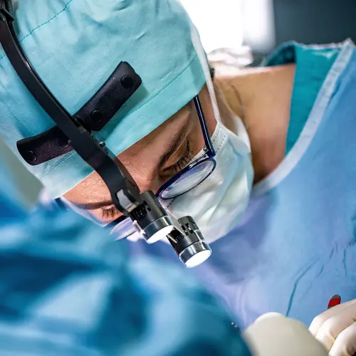
Heart surgery. The defect may be very large or located in such a place that a patch cannot be placed. Open-heart surgery is needed for treatment of such ASDs. Your child will be unconscious during the operation. The surgeon will open the heart and sew a patch over the ASD. If the ASD is small, they may simply stitch it closed. The hospital stay will be several days, and the chest incision needs six weeks to heal.
Primum and sinus venosus type ASDs usually necessitate open-heart surgery.
ASD surgery has high success rates. There are no long-term problems once the chest incision and heart have healed.
Most children born with ASD grow up to live a normal, active life.

